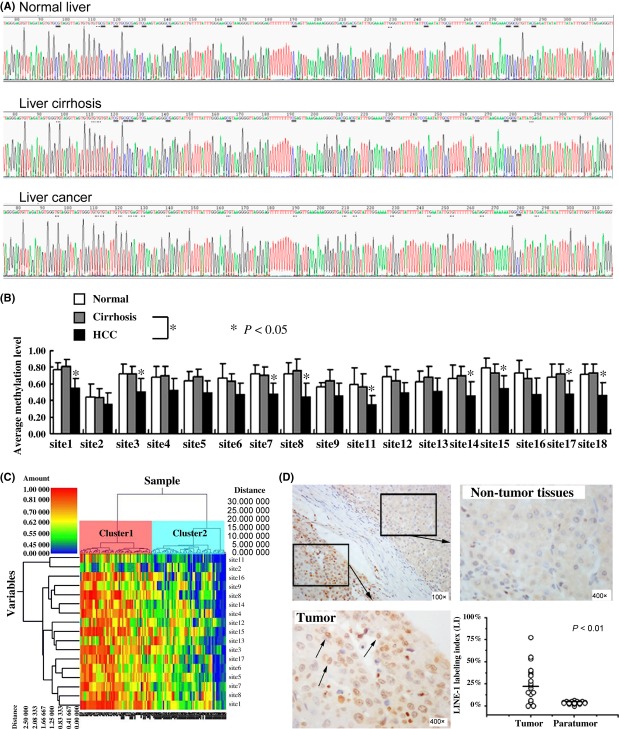
Figure 2.
Methylation Level of long interspersed nucleotide element-1(LINE-1) promoter and expression of LINE-1 in liver tissue. (A) Representative bisulfite sequencing analysis of the LINE-1 promoter in normal liver, cirrhosis and liver cancer. The methylated and unmethylated CpG sites corresponding to the 19 CpG sites are underlined in parallel, methylated CpGs were CG, whereas unmethylated CpGs were TG. (B) Methylation levels of all CpG sites in LINE-1 promoter were compared between samples from subjects with HCC or cirrhosis and normal liver control. *P < 0.05, relative to the respective control. (C) HeatMap showing unsupervised clustering of normal, cirrhosis and HCC samples (x-axis) between the methylation of all CpG (y-axis). Green corresponds to low methylation and Red to high methylation. (D) Representative samples of immunostaining for LINE-1 and quantitative analysis results for LINE-1-positive dots in HCC and corresponding non-tumourous liver tissues. LINE-1 staining in HCC showed nuclear positivity (arrows), but negative in non-tumourous liver tissues (× 400).

