
Figure 1.
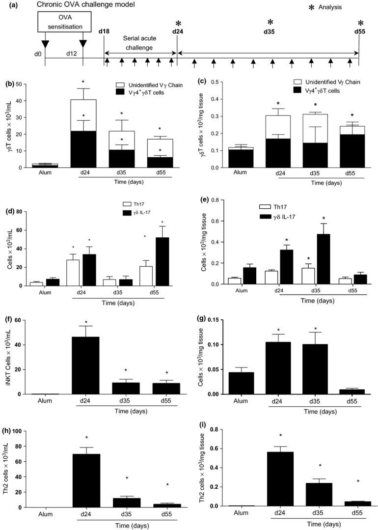
Pulmonary γδT cells are elevated following allergen exposure. Schematic OVA challenge model (a). Animals were systemically sensitised with alum/OVA (alum/PBS) followed by acute OVA challenge (days 18–24). To induce chronic inflammation and remodelling, OVA challenges were continued 3× weekly. Mice were sacrificed 24 h after final aerosol challenge on days 24, 35 and 55. Total and Vγ4+ γδT cells in the BAL (b) and lung (c). Th17 and IL-17+ γδT cells in the BAL (d) and lung (e). iNKT cells in the BAL (f) and lung (g). Th2 cells in the BAL (h) and lung (i). Data represent mean ± SEM (n = 6–12). *P < 0.05 compared to alum controls, (Mann–Whitney U-test). Alum controls from days 24, 35 and 55 were not significantly different between time points and were pooled for clarity.