
Fig. 1. Design of DNA-brick crystals.
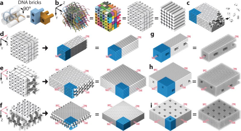
a, A strand (left) and a brick (right) model showing two 32-nt DNA bricks that form a 90° angle. b, Models of a 6H (helix) × 6H (helix) × 24B (basepair) cuboid with increasing levels of abstraction: a strand (leftmost) and a brick model where colors distinguish brick species, a brick model with all bricks colored grey, and a model where cylinders representing DNA double-helices. c, Individual DNA strands, rather than pre-assembled multi-brick blocks, are directly incorporated into the growing crystal. d to f, Brick and cylinder models of a 1D Z-crystal (d), a 2D ZX-crystal (e), and a 2D XY-crystal (f) designed from the 6H×6H×24B cuboid. g to i, Cylinder and DNA-helix models of crystals with pores and tunnels. g, A Z-crystal with a tunnel and periodic pores. h, A ZX-crystal with two groups of parallel tunnels. i, An XY-crystal with periodic pores. Repeating units of the crystals are denoted using blue-colored boxes. Pink arrows indicate the directions of crystal growth.