
Fig. 2. One-dimensional DNA crystals.
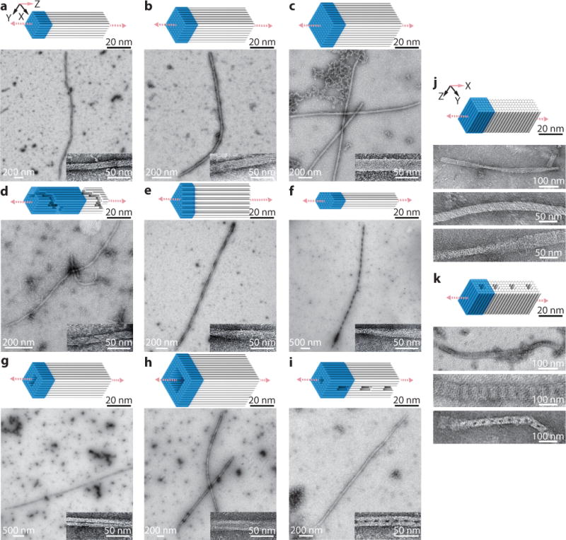
a to i, Z-crystals with cylinder models and TEM images. a to c, Z-crystals with solid cross-sections: 6H×6H (a), 8H×8H (b), and 10H×10H (c) Z-crystals. d to f, Z-crystals with different cross-sectional shapes: an 8H×8H Z-crystal with right-handed spiral channel (d), a 43H Z-crystal with triangle-shaped cross-section (e), and a 44H Z-crystal with hexagon-shaped cross-section (f). g to i, Z-crystals with porous cross-sections: an 8H×8H Z-crystal with a 2H×4H tunnel (g), a 12H×12H Z-crystal with a 6H×6H tunnel (h), and an 8H×8H Z-crystal with a 2H×2H tunnel and perpendicular 8H×2H×24B pores (i). j and k, cylinder models (top) and TEM images (bottom) of X-crystals. j, an X-6H×6H×64B-cuboid crystal. k, a 6H×6H X-crystal with 2H×2H pores. Unit cells of crystals are denoted using blue-colored boxes. See Supplementary Figs. S3 to S12 for more TEM images.