
Fig. 3. Two-dimensional multilayer ZX-crystals.
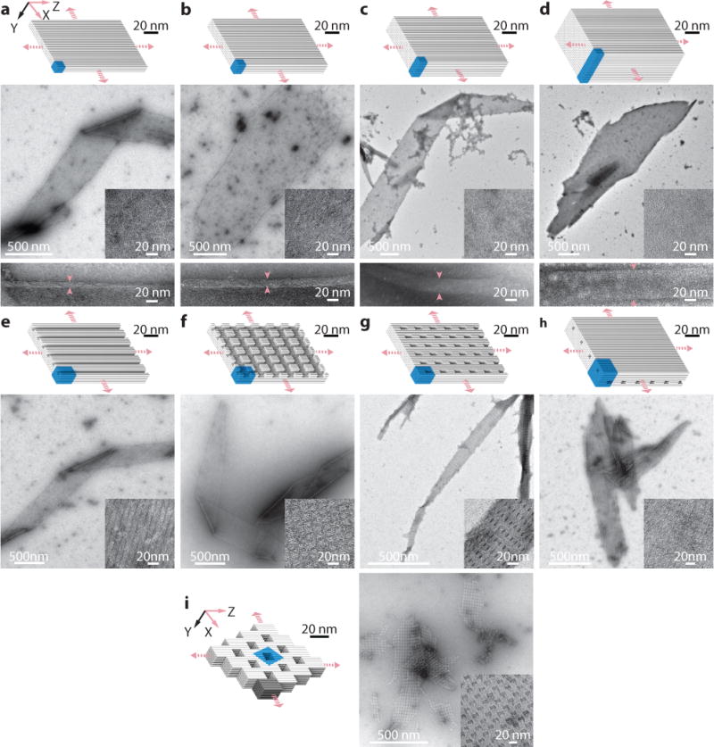
Cylinder models (top) and TEM images (bottom) are shown for each crystal. a to d, Solid ZX-crystals: 4-layer (a), 6-layer (b), 10-layer (c), and 20-layer (d), solid ZX-crystals. Arrows indicate the positions for the thickness measurement of the crystals. e to h, ZX-crystals with channels, pores, and tunnels: 6-layer crystal with 2H×2H parallel channels (e), 6-layer crystal with two groups of crossing channels – 2H×2H channels that run parallel to the DNA helical axis and 2H×32B channels that run perpendicular to the DNA helical axis (f), 6 layer crystal with 2H×6H×32B pores (g), and 10 layer crystal with two groups of non-contacting tunnels – 2H×2H tunnels parallel to the DNA helical axis and 2H×24B tunnels perpendicular to DNA helical axis (h). In h, the two groups of tunnels are separated by two layer of DNA helices. i, An offset-ZX-6H×6H×64B-cuboid ZX-crystal. The dark grey part represents a 6H×6H×64B-cuboid repeating unit. Unit cells of crystals are denoted using blue-colored boxes. See Supplementary Figs. S13 to S21 for more TEM images.