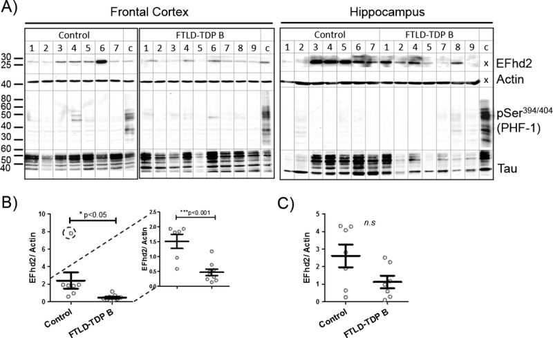
FIGURE 3.

EFhd2 protein levels in the frontal cortex are reduced in patients with dementia without tauopathy. (A) Western blots showing EFhd2 protein levels in human frontal cortex (left) and hippocampal (right) tissue RIPA extracts from nondemented controls and individuals with frontotemporal lobar degeneration FTLD-TDP Type B. A sample from AD frontal cortex was run as a calibrator and phospho-tau–positive control on each gel. No calibrator sample was run for EFhd2 and actin detection in hippocampal samples because all samples were run on the same SDS-PAGE. Beta-actin was used as a loading control. (B) Quantification of EFhd2 protein bands by densitometry in frontal cortex samples. * p < 0.05 with all data points (left), *** p < 0.001 without encircled outlier (right), compared with control by t test. (C) Quantification of EFhd2 protein bands in samples from human hippocampus from nondemented controls and individuals with FTLD-TDP Type B.
