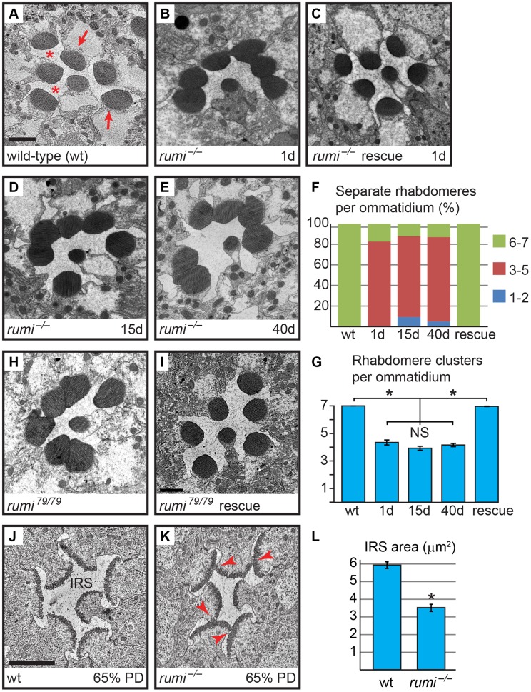
Figure 1. Loss of the enzymatic function of Rumi results in rhabdomere adhesion.
Shown are electron micrographs of a single ommatidium from adult (A–E,H,I) or 65% PD (J,K) from the indicated genotypes. All animals were raised at 18°C. (A) Wild-type. Arrows indicate rhabdomeres and asterisks indicate the IRS. Scale bar: 2 µm. (B) 1-day old rumi−/−. Note the attachment in the neighboring rhabdomeres. (C) 1-day old rumi−/− expressing a wild-type P{rumigt-FLAG} genomic transgene. (D,E) The rumi−/− rhabdomere attachment phenotype does not change with age, since 15-day old (D) and 40-day old (E) animals show a similar degree of attachment. (F) Percentage of number of individual rhabdomeres per ommatidia for various genotypes. At least three animals were used for each genotype. The number of ommatidia examined for each genotype is as follows: wt (50), 1d (35), 15d (66), 40d (85), rescue (126). (G) Quantification of average individual rhabdomere number per ommatidium for the data shown in F. Rhabdomere attachments in 1-day, 15-day and 40-day old rumi animals are not significantly different from one another, but are significantly different from wild-type and rescued animals (*P<0.0001). NS, not significant. (H,I) The enzymatically inactive allele rumi79 also shows a rhabdomere attachment phenotype (H), which can be rescued by one copy of the wild-type P{rumigt-FLAG} genomic transgene (I). (J,K) Ommatidia from animals at 65% PD from wild-type (J) and rumi−/− (K). Arrowheads in (K) indicate points of rhabdomere attachment. (L) Means ± SEM of IRS area in wild-type and rumi−/− at 65% PD. IRS areas were measured using ImageJ software. Unpaired t-test was used to compare wt (n = 14) and rumi−/− (n = 24) IRS, *P<0.0001.

