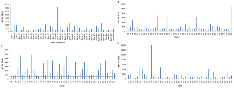
Figure 1. Detection of NiVsG antibodies in Luminex based multiplexed microsphere assay.
The median fluorescent intensities (MFI) for each microsphere population are shown in graphs. MFI for antibody positive cattle and goat shown in graph I. MFI for antibody positive pig sera is shown in graph II, III and IV. The gray bar represents the detection cut-off of 300 MFI for cattle and goat sera and 650 MFI for pig sera.

