
Figure 7.
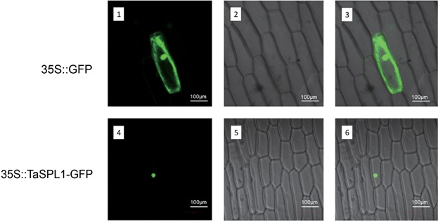
Subcellular localization of TaSPL17–green fluorescent protein (GFP) fusion proteins driven by the CaMV35S promoter The vector control (35S::GFP) and the fusion proteins (35S::TaSPL17–GFP) were introduced into onion epidermal cells and observed with a laser scanning confocal microscope. Images are in dark field (1, 4), bright field (2, 5), and combined (3, 6).