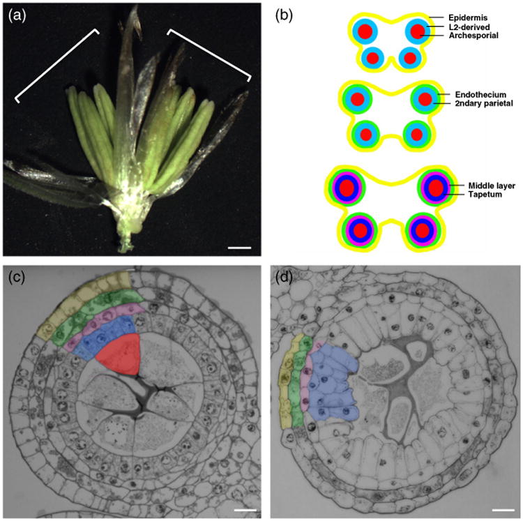
Figure 1.

Anther development in maize.
(a) Maize spikelet showing the two florets (white brackets) with three anthers each. Scale bar: 1 mm.
(b) Schematic drawing of anther development in maize.
(c, d) Transverse section of fertile (c) and ms32-ref (d) anthers. Layers in (c) are color coded as in (b). Same colors are used for the middle layer and tapetal precursor cells in (d). Scale bars: 20 μm.
