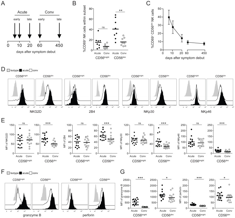
Figure 1. CD56dim NK cells are highly activated in HFRS patients.
(A) PBMC from 16 PUUV-infected HFRS patients were collected in the early acute (median d6) and convalescence phase (d60). From 8 HFRS patients additional samples were collected at the indicated time-points and up to day 450. Analyses of NK cell phenotype shown in 1B and 1D–G were performed with samples from patients in the early acute (median d6) and early convalescent phase (d60) of HFRS. (B) Frequencies of CD69-positive CD56bright and CD56dim NK cells in early acute and convalescent (d60) HFRS infection (n = 8). (** p≤0.01, paired t-test). (C) Frequencies of CD69-positive CD56dim NK cells from HFRS patients (n = 8) from early acute to convalescence phases are depicted. Values shown are mean (+/− SD). (D and E) Expression levels of activating NK cell receptors on CD56bright and CD56dim NK cells in acute and convalescent phases (D) Representative staining for NKG2D, 2B4, NKp30 and NKp46 on CD56bright and CD56dim NK cells during acute HFRS infection (black) and convalescence (white). Isotype (grey). (E) Expression levels (MFI) of NKG2D, 2B4, NKp30 and NKp46 on CD56bright and CD56dim NK cells in HFRS patients (n = 14–16) in acute (black) and convalescent phases (white). (*** p≤0.001, paired t-test). (F and G) Levels of intracellular cytotoxic effector molecules in CD56bright and CD56dim NK cells in acute and convalescent phases. (F) Representative intracellular staining for granzyme B and perforin in CD56bright and CD56dim NK cells in acute HFRS infection (black) and convalescence (white). Isotype (grey). (G) Expression levels (MFI) of intracellular granzyme B and perforin in CD56bright and CD56dim NK cells of HFRS patients (n = 14–16) in acute (black) and convalescent phases of infection (white). (*** p≤0.001, * p≤0.05, paired t-test).

