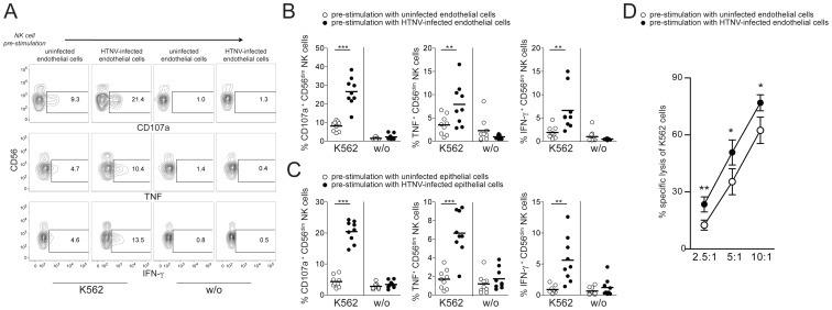
Figure 4. CD56dim NK cells acquire increased functional capacity after contact with hantavirus-infected cells.
(A–C) Degranulation (CD107a) and intracellular cytokine production of CD56dim NK cells reacting to K562 cells after pre-stimulation with uninfected or HTNV-infected endothelial and epithelial cells. (A) Representative FACS analysis of CD107a, IFN-γ and TNF expression in one NK cell donor is shown. (B and C) Summary of the CD56dim NK cell responses against K562 cells (n = 9) after pre-stimulation with uninfected (white) or HTNV-infected (black) endothelial (B) and epithelial cells (C). (*** p≤0.001, ** p≤0.01; paired t-test). (D) NK cell-mediated specific lysis of K562 cells after pre-stimulation of NK cells with uninfected (white) and HTNV-infected (black) endothelial cells. Depicted are mean values (+/− SD) from 5 donors and 2 independent experiments (** p≤0.01, *p≤0.05; paired t-test).

