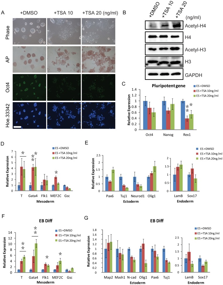
Figure 1. TSA induces early differentiation of ESCs and promotes mesodermal lineage differentiation.
(A) Bright-field images, alkaline phosphatase staining of ESCs and representative immunofluorescence images of Oct4 staining in control or TSA-treated ESCs (10 and 20 ng/ml) in the presence of LIF. (B) Western blotting verification of H3, acetyl-H3, H4, and acetyl-H4 in control or TSA-treated ESCs (10 and 20 ng/ml). GAPDH was used as a loading control. (C) The relative expression levels of Oct4, Nanog, and Rex1 mRNA in control or TSA-treated ESCs (10 and 20 ng/ml). (D, E) QRT-PCR analysis for marker genes of three germ layers (endoderm, mesoderm and ectoderm) in control or TSA-treated ESCs (10 and 20 ng/ml), under the monolayer differentiation condition without LIF. The cells were treated by TSA after removing LIF for 24h and collected mRNA for QRT-PCR analysis at day 3 of monolayer differentiation. (F, G) QRT-PCR analysis for marker genes of the three germ layers in control or TSA-treated ESCs (10 and 20 ng/ml) during EB differentiation. The EBs was treated by TSA from day 2 to 6 of EB differentiation. Data are expressed as means ± SD. Statistical significance was assessed by two-tailed Student's t test. ***, P<0.001; **, P<0.01; *, P<0.05.

