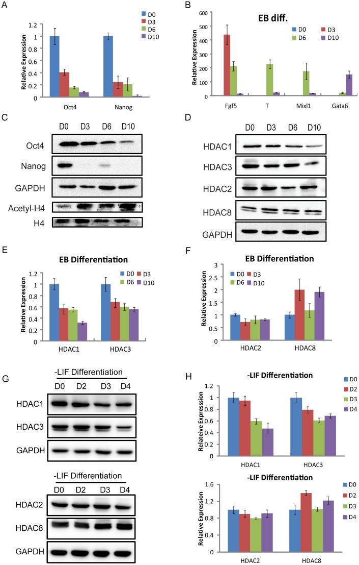
Figure 2. The expression levels of HDAC1 and 3 are decreased during differentiation.
(A) QRT-PCR for genes characteristic of undifferentiated stem cells (Oct4, Nanog) was performed as indicated on mRNA collected at days 0, 3, 6, and 10 during EB differentiation. (B) The relative expression levels of marker genes for three germ layers (endoderm, Gata6; mesoderm, T, Mixl1; primitive ectoderm, Fgf5) at days 0, 3, 6, and 10 during EB differentiation. (C) Western blotting verification for genes characteristic of undifferentiated stem cells (Oct4, Nanog) was performed as indicated on protein samples collected at days 0, 3, 6, and 10 during EB differentiation. The expression level of global acetyl-H4 was increasing during EB differentiation. GAPDH and H4 were used as loading controls. (D) Western blotting verification for class I HDAC members (HDAC1, 2, 3, and 8) at the indicated days 0, 3, 6, and 10 during EB differentiation. GAPDH was used as a loading control. (E, F) QRT-PCR analysis for the expression levels of class I HDAC members (HDAC1, 2, 3, and 8) at the indicated days 0, 3, 6, and 10 during EB differentiation. (G, H) Western blotting and QRT-PCR analysis for the expression levels of HDAC members (HDAC1, 2, 3, and 8) at days 0, 2, 3, and 4 during differentiation without LIF.

