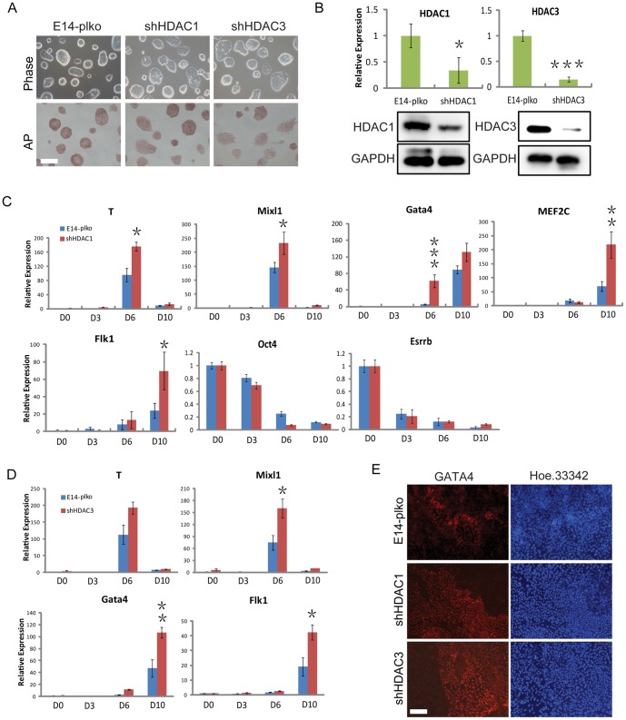
Figure 3. Loss of HDAC1 or 3 enhances mesodermal lineage differentiation.
(A) Bright-field images and alkaline phosphatase staining of ESCs in shHDAC1 and shHDAC3 ESCs. (B) Western blotting verification and QRT-PCR analysis of the knockdown of HDAC1 and HDAC3 in stable E14 cell lines. GAPDH was used as a loading control. (C) QRT-PCR analysis of mesoderm genes in shHDAC1 ESCs and control cells at the days 0, 3, 6, and 10 during EB differentiation. (D) QRT-PCR analysis of mesoderm genes in shHDAC3 ESCs and control cells during EB differentiation. (E) Representative immunofluorescence images for the GATA4 expression level in control, shHDAC1, and shHDAC3 cells after 9 days of EB formation. Green, Gata4; blue, Hoechst 33342 for nuclei staining. Data are expressed as means ± SD. Statistical significance was assessed by two-tailed Student's t test. ***, P<0.001; **, P<0.01; *, P<0.05.

