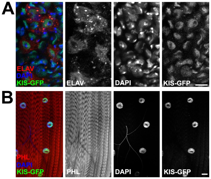
Figure 1. Kis localizes to the nucleus of motor neurons and muscles.
(A) Confocal images of Kis-GFP expression in midline of third instar larval ventral nerve cord expressing RFP in all neurons using the Elavc155-Gal4 driver. Neurons are labeled in red (Elav), nuclei in blue (DAPI), and Kis in green (Kis-GFP). Note presence of Kis-GFP in neuron nuclei. Right panels show individual channels. Scale bar = 10 µm. (B) Confocal images of Kis-GFP expression in multi-nucleated muscle cells of muscles 6 and 7 in third instar larval NMJs. Muscles are labeled with phalloidin (PHL, red), muscle nuclei in blue (DAPI), and Kis in green (Kis-GFP). Note presence of Kis-GFP in muscle nuclei. Right panels show individual channels. Scale bar = 50 µm.

