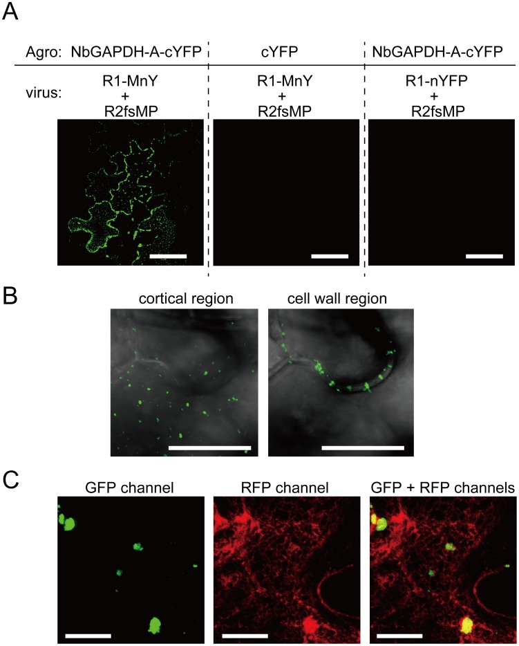
Figure 7. Bimolecular fluorescence complementation analyses of the interaction between RCNMV MP and NbGAPDH-A.
NbGAPDH-A fused to the C-terminal half of YFP at the C-terminus (NbGAPDH-A-cYFP), or C-terminal half of YFP (cYFP) as the negative control, was expressed with TBSV silencing suppressor p19 in N. benthamiana leaves via Agrobacterium infiltration. 18 h after infiltration, in vitro transcripts of the recombinant RCNMV that expressed fusion protein of the MP and N-terminal half of YFP at the C-terminus (R1-MnY+R2fsMP, Figure S1I), or the recombinant virus that expressed N-terminal half of YFP (R1-nYFP+R2fsMP, Figure S1J) as the negative control, was mechanically inoculated. At 28 hpi of the recombinant virus, reconstructed YFP signal was visualized using CLSM. (A) Reconstituted YFP signals were detected as foci composed of 5–10 cells in the leaves that expressed NbGAPDH-A-cYFP and inoculated with R1-MnY+R2fsMP (left panel). No YFP signals were detected in the leaves that expressed unfusedcYFP (center panel), or those inoculated with R1-nYFP+R2fsMP (right panel). Scale bar = 50 µm (B) Large magnification images of the reconstituted YFP in the cortical (left panel) and inner cell wall region (right panel). Images are from optical sections taken at upper part for cortical region and middle part for cell wall region in the same site of the leaf and mergers of DIC and GFP channel. Scale bar = 10 µm (C) Reconstituted YFP signals as cortical punctates (left panel), ER-mCherry signals (center panel) and overlapped (right panel). Most YFP-punctates are larger than those in (B), because this cell is closer to the center of infection and that shows the cell is at a later stage of virus infection. Scale bar = 10 µm.

