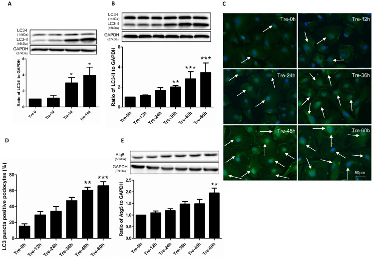
Figure 1. Trehalose induced autophagy in human podocytes.
(A) The expression of LC3-II increased in a dosage dependent manner. Conditionally immortalized human podocytes were treated with 0, 10, 50 and 100 mM of trehalose (Tre) for 48 h. LC3-II was measured by Western blotting. The data (means ± SEM) was expressed as the relative changes compared with Tre-0 mM group. Representative immunoblot images were shown along with the statistical results. *p<0.05 versus Tre-0 mM, n = 5. (B) LC3-II increased in a time dependent manner. Podocytes were treated with 50 mM Trehalose for 0, 12, 24, 36, 48 and 60 h. **p<0.01, ***p<0.001 versus Tre-0 h, n = 7. (C–D) LC3-II puncta increased after trehalose treatment. LC3 immunostaining in podocytes was performed at 0, 12, 24, 36, 48 and 60 h after trehalose treatment (50 mM). Significant increased green bright puncta (indicated by white arrows) can be observed in cytoplasm after 48 h-trehalose treatment. The representative LC3 immunostaining images were shown along with statistical results from 6 independent experiments. **p<0.01, ***p<0.001 versus Tre-0 h. Podocyte nuclei were stained with DAPI (blue). (E) The expression of Atg5 was up-regulated in trehalose-treated podocytes (50 mM). Podocytes were treated with 50 mM Trehalose for 0, 12, 24, 36, 48 and 60 h. The expression of Atg5 significantly increased at the time point of 60 h. **p<0.01 versus Tre-0 h, n = 5.

