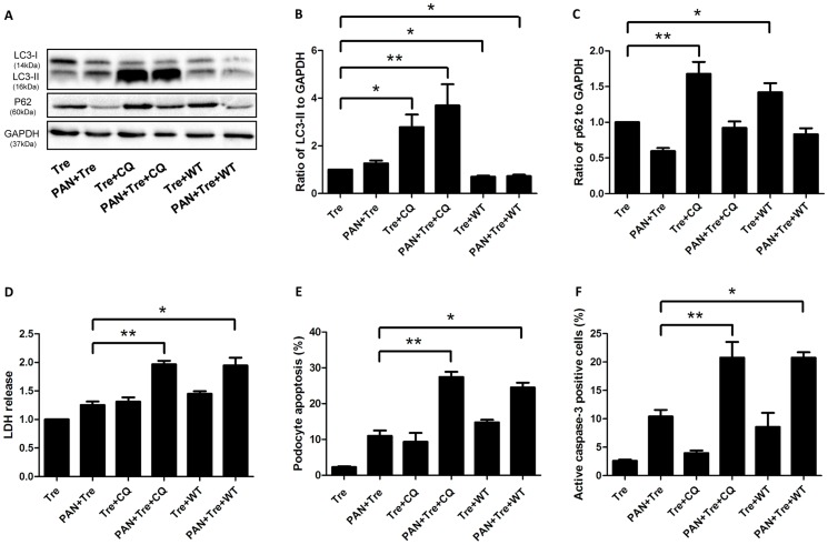
Figure 5. Inhibition of trehalose-induced autophagy abolished its cytoprotective effects in preventing podocyte apoptosis.
CQ (25 µM) or WT (0.2 µM) was used to inhibit podocyte autophagy which was induced by trehalose (50 mM) or trehalose (50 mM) + PAN (30 µg/ml) for 48 h. (A–C) CQ and WT inhibited trehalose-induced autophagy. The expression of LC3-II drastically increased in Tre+CQ and PAN+ Tre+CQ groups, while it decreased significantly in Tre+WT and PAN+ Tre+WT groups. p62 slightly decreased in PAN+ Tre group, whereas it significantly increased in Tre+CQ and Tre+WT groups. The immunoblot images were shown along with statistical data. *p<0.05, **p<0.01 versus Tre group, n = 7. (D) Necrosis increased after the inhibition of trehalose-induced autophagy. The LDH in culture medium was measured. *p<0.05, **p<0.01 versus PAN+Tre group, n = 6. (E) Podocyte apoptosis increased after the inhibition of trehalose-induced autophagy. The percentage of apoptotic podocytes was much higher in PAN+Tre+CQ and PAN+Tre+WT groups than the PAN+ Tre group. *p<0.05, **p<0.01 versus PAN+ Tre group, n = 7. (F) The percentage of active caspase-3 positive podocytes increased after inhibition of trehalose-induced autophagy. The changes in the percentage of active caspase-3 positive podocytes were similar to the data in (E). *p<0.05, **p<0.01 versus PAN+ Tre group, n = 8.

