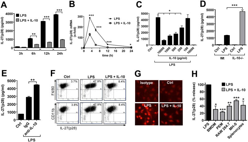
Figure 4. Suppression of IL-27(p28) production by IL-10 in macrophages.
A. Time course of IL-27(p28) release from BMDM (Wt) after LPS (1 μg/ml) +/- IL-10 (10 ng/ml), ELISA. B. RT-PCR of IL-27(p28) mRNA levels in BMDM (Wt) with LPS or LPS plus IL-10 (10 ng/ml). C. Dose response studies of IL-27(p28) release from LPS-activated BMDM (Wt) with several IL-10 concentrations, 10 h. Ctrl indicates resting BMDM. D. Release of IL-27(p28) by LPS-activated BMDM from Wt or IL-10-/- mice, 10 h. E. Release of IL-27(p28) by BMDM (Wt) after LPS with addition of control IgG or neutralizing anti-IL-10 antibody (10 μg/ml), 24 h. F. Flow cytometry of BMDM (Wt) after LPS +/- IL-10, 10 h. G. Immuno-cytofluorescence of BMDM (Wt) with red staining for IL-27(p28) (Olympus BX-51 microscope [100×/1.4 oil] with Olympus DP-70 camera), 14 h. H. Relative inhibition of IL-27(p28) by IL-10 in BMDM, peritoneal elicited macrophages (PEM), RAW 264.7 macrophages, MH-S macrophages (SV40 transformed mouse alveolar macrophage cell line) and splenocytes from C57BL/6J mice, 10 h. Data are representative of three independent experiments each performed in duplicates. * P < 0.05, ** P < 0.01, *** P < 0.001.

