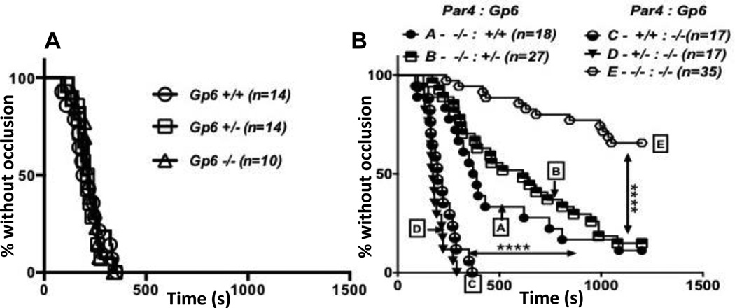
Figure 5. Effect of combined partial or complete Par4 and GPVI deficiency on carotid artery occlusion.
Carotid arteries of anesthetized 8 to 14 week-old mice were injured by exposure to filter paper saturated with 1.25 M (20%) FeCL3, flow after injury was measured, and time to stable occlusion determined. Data are shown as percent of arteries without occlusion as a function of time after injury for each genotype. A) Littermate offspring of Gp6+/− crosses were examined. B) Littermate offspring of Par4+/−:Gp6+/− crosses and Par4−/−:Gp6+/−×Par4−/−:Gp6−/− crosses were examined. Note that GPVI deficiency alone had no effect. In (B), genotype affected time to occlusion by log-rank analysis (P<0.0001). In multiple comparison-corrected follow-on comparisons, Par4 deficiency alone prolonged time to occlusion (curve A vs. C,D; P<0.0001), and combined Par4 and GPVI deficiency provided a substantial further increase in time to occlusion (curve E vs. A; P<0.0001).

