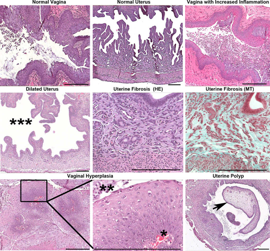
Fig. 5.
Representative images from histopathologic analysis of the uterus and vagina. Treatment of AdCre-infected wild-type or LSL-K-RasG12D/+ PtenloxP/loxP mice with nanoparticles did not result in pathologic changes beyond those seen in untreated AdCre-infected mice. Pathologic conditions observed after infection with AdCre included: increased vaginal inflammation, uterine dilation (***), and uterine fibrosis. Uterine fibrosis was confirmed by the presence of collagen (blue) by Masson’s trichrome (MT) staining. A single LSL-K-RasG12D/+ PtenloxP/loxP mouse infected with AdCre and treated with NP (CPT) had a focus of squamous hyperplasia with orderly progression of squamous epithelial cells from the basement membrane (*) to the surface (**) in the vagina. Additionally, a single LSL-K-RasG12D/+ mouse infected with AdCre had a uterine polyp (arrow). Scale bars 200 µm

