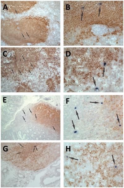
Figure 1. Localization of SIV RNA+ cells in secondary lymphoid tissues of chronically infected rhesus macaques.
Representative images of in situ hybridization for SIV RNA to identify virus-producing cells (blue/black cells indicated by arrows) and CD20 staining (brown) to morphologically identify B cell follicles in spleen (A, B), axillary lymph node (C, D), ileum (E, F) and colon (G, H). Images B, D, F and H are high magnification (original magnification 252X) images from the fields shown in A, C, E and F (original magnification 63X), respectively.

