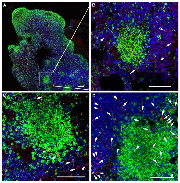
Figure 5. Localization of SIV-specific CTL in secondary lymphoid tissues of rhesus macaques during chronic infection.
Representative lymph node tissue sections stained with MHC class I tetramers (red) to label SIV-specific CTL, CD3 antibodies (blue) to label T cells, and CD20 antibodies (green) to label B cells and delineate B cell follicles. (A) Shows a montage of multiple confocal projected z-scans from an inguinal lymph node from animal Rhau10 stained with Mamu-B*008:01/Vif RL8 tetramers. (B-D) are confocal z-scans showing the range of localization patterns of SIV-specific CTL within follicles typically seen in spleen and lymph nodes from animals in the study. (B) Enlargement from A demonstrating a B cell follicle devoid of CTL. (C) Tissue section from the same lymph node shown in A and B now stained with Mamu-B*008:01/Vif RL9 tetramers demonstrating an example of CTL located on the follicle edge. (D) A mesocolonic lymph node section from animal R03116 stained with Mamu-A*001:01/Gag CM9 tetramers exemplifying CTL distributed throughout a follicle. Tetramer+ cells within the sections were identified in montages of high-resolution serial z-scans, and are indicated by arrows in B-D. Within individual z-scans, tops and bottoms of cells were distinguished from non-specific background staining by stepping up and down through the adjacent z-scans. Red staining in the images without arrows was determined to be background staining by this technique. Confocal images were collected with a 20X objective and each scale bar indicates 100 μm.

