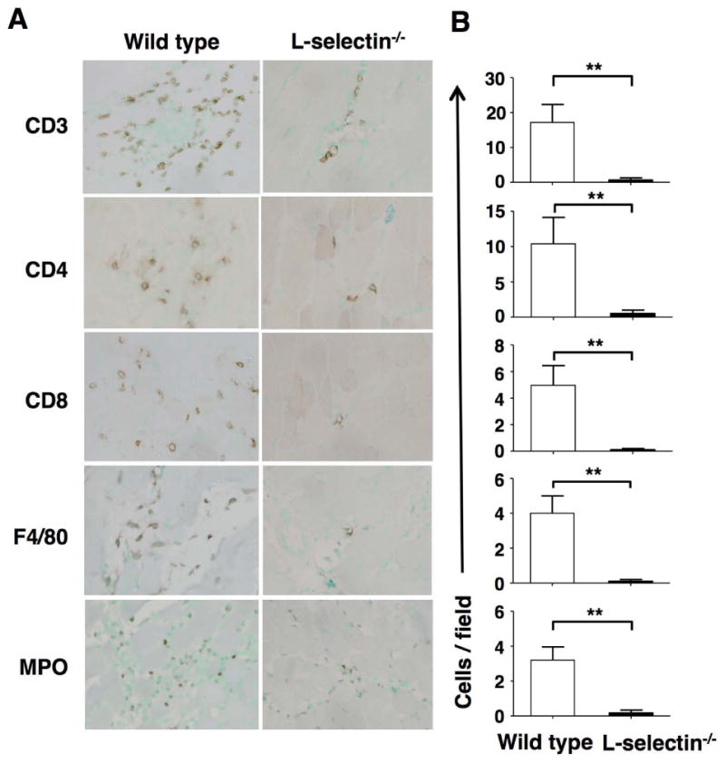
Figure 2.

Inflammatory cell infiltration into inflamed muscles of wild type (WT) and L-selectin-/- mice during C protein-induced myositis. Mice were sacrificed at day 14 after immunization with C protein and muscle tissues were harvested. A, Representative immunohistochemical images showing CD3, CD4, CD8, F4/80, and myeloperoxidase (MPO) staining in tissue sections from L-selectin-/- mice and wild type mice. B, Numbers of CD3+, CD4+, and CD8+ T cells, F4/80+ macrophages, and MPO+ neutrophils on day 14. Bars show the mean ± SEM. These results represent those obtained with 8-10 mice of each genotype. **p < 0.01. A, Original magnification: ×400.
