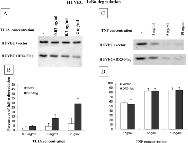
Figure 3.

TL1A induced IκBα degradation in HUVEC. (A) HUVEC transfected with mock vector or DR3 were treated with ascending concentration of TL1A for 30 minutes, IκBα showed a clear degradation after TL1A stimulation for the DR3-Flag transfected EC. Blots were representatives of 3 separate experiments. (B) Densitometry analysis showed a significant increase in IκBα degradation by TL1A at 0.2 and 2 μg/ml for the DR3 transfected EC compared to the mock transfected (13.3 ± 4 vs.4.3 ± 3.2, 29 ± 5.5 vs.7.2 ± 5.6 respectively) (p < 0.05). Values were expressed as percentages of reduction compared to TL1A 0 μg/ml IκBα densities. Results were mean ± SE from 3 experiments. (C, D) There was no difference in IκBα degradation after TNF treatment between HUVEC transfected with empty vector and DR3.
