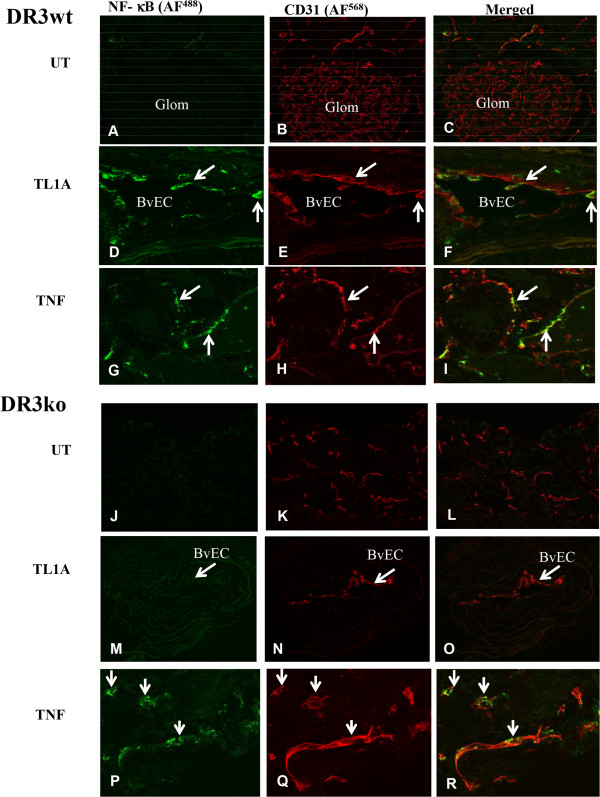
Figure 5.

TL1a induced NF-κB activation in renal interstitial vascular endothelial cells in mouse organ cultures. Kidney tissue from DR3 wild type (wt) and DR3 knockout (ko) mice were treated with TL1A or TNF and immunolabeled as described in materials and methods. (A-C) Untreated (UT) DR3wt showed no positive staining for NF-κB activation in CD31 positive EC from glomerulus or interstitial vessels. (D-F) some interstitial EC were positive for NF-κB activation after TL1A treatment. (G-I) more vascular EC were positive for NF-κB activation after TNF treatment. (J-L) untreated DR3ko showed no staining for NF-κB activation in vascular EC. (M-O) TL1A-treated DR3ko showed no staining for NF-κB activation in vascular EC. (P-R) TNF-treated DR3ko showed positive staining for NF-κB activation in vascular EC. Arrow for BvEC: blood vessel endothelial cells. Results were representative of 3 experiments.
