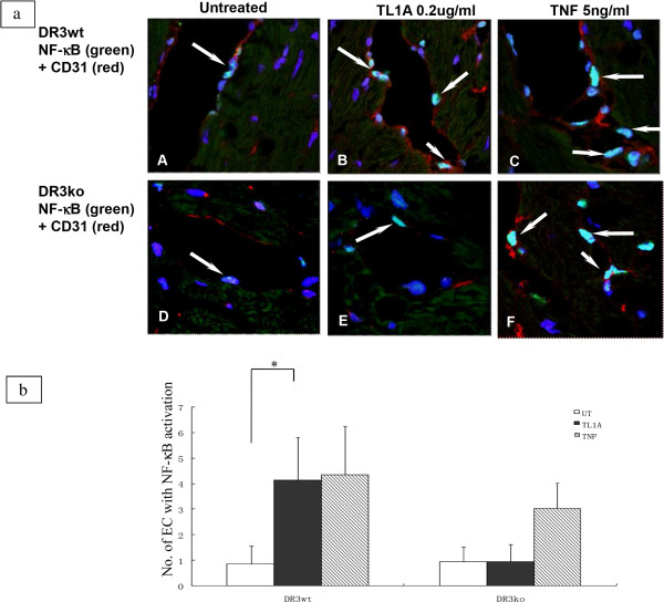
Figure 6.

TL1A induced NF-κB activation in vascular endothelial cells in mouse heart organ cultures. (6a) Heart tissue from DR3 wild type (wt) and DR3 knock-out (ko) mice were treated with TL1A or TNF and immunolabeled as described in materials and methods. DR3wt showed more nuclear NF-κB activation in CD31 positive EC after TL1A treatment (B) compared to the untreated (A). There was similar odd nuclear NF-κB activation in DR3ko mice EC between TL1A treated (E) and untreated cultures (D). TNF induced a more profound nuclear NF-κB activation in EC of both DR3wt and DR3ko (C and F). Results were representative of 3 experiments. (6b) Graph showed a statistical difference in number of EC in TL1A treated tissue with NF-κB activation in DR3wt compared to DR3ko (p < 0.05) (*).
