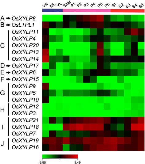
Figure 4.

Expression profiles of OsXYLP genes in various organs and tissues. The microarray data (GSE6893) of OsXYLP genes expression are analyzed. A heat map representing hierarchical clustering of average log signal values of OsXYLP genes in various developmental stages are generated (samples are indicated at the top of each lane: YR, roots from 7-day-old seedlings; ML, mature leaves; YL, leaves from 7-day-old seedling, different stages of panicle development: SAM, up to 0.5 mm; P1, 0–3 cm; P2, 3–5 cm; P3, 5–10 cm; P4, 10–15 cm; P5, 15–22 cm; P6, 22–30 cm and different stages of seed development: S1, 0–2 dap (days after pollination); S2, 3–4 dap; S3, 5–10 dap; S4, 11–20 dap; S5, 21–29 dap). Genes are divided into 10 groups: (A) SAM, P1-P6, S1-S5; (B) all examined organs and tissues; (C) YR, P4-P6; (D) ML, P5, P6; (E) YR, P4-P6; (F) P3; (G) YR; (H) low expression in all examined organs and tissues; (I) SAM, P1-P6, S3-S5; (J) all examined organs and tissues. The color scale (representing average log signal values) is shown at the bottom.
