
Figure 2.
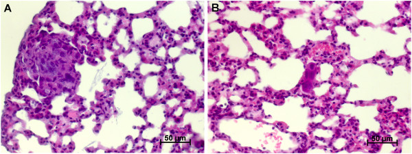
Typical metastases in pfg / rag2 and rag2 mice. A) Typical metastasis of pfp/rag 2 mice with 10–100 malignant cells forming the metastatic deposit. B) Typical metastasis of rag2 mice with only very few malignant cells. Magnification: 400X, staining method: haematoxylin and eosin (H.E.).