
Figure 1. The GAP Activity of RGA-3/4 restrains RhoA during mitosis and cytokinesis.
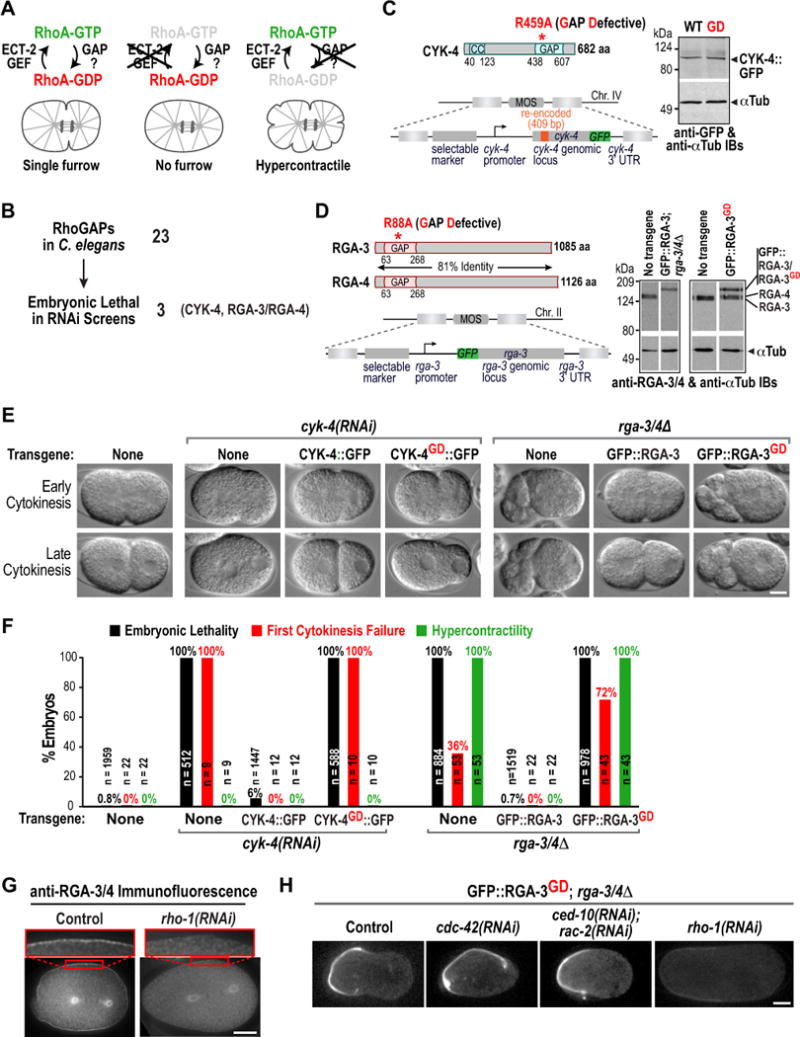
(A) Schematics of a wild-type embryo (left), an ECT-2 depleted embryo (middle); and of the predicted effect of inhibiting the RhoA GAP (right). (B) Of the 23 RhoGAPs in C. elegans only three (CYK-4, RGA-3, and RGA-4) are embryonic lethal (Table S1). (C) (left) Schematics of CYK-4 and the cyk-4::gfp transgene. (right) Immunoblot of worm extracts from the indicated transgenic strains probed for GFP and α-tubulin as a loading control. (D) (left) Schematics of RGA-3 and RGA-4 and of the gfp::rga-3 transgene. (right) Immunoblot of worm extracts from N2 control and indicated transgenic strains; the strain expressing GFP::RGA-3 was homozygous for rga-3 and rga-4 deletions (rga-3/4Δ), whereas the strain expressing GFP::RGA-3GD was not. (E) DIC images during early and late cytokinesis for the indicated conditions. (F) Graph plotting embryonic lethality, first division cytokinesis failure, and hypercontractility (>2 ingressions during mitosis). n = number of embryos. (G) Immunofluorescence of one-cell stage wild-type (left) and rho-1(RNAi) (right) embryos stained with antibodies to RGA-3/4 (n>12 for each condition). (H) Central plane confocal images of one-cell stage metaphase rga-3/4Δ embryos expressing GFP-RGA-3GD after RNAi of the indicated genes (n>7 embryos for each condition). Bars, 10μm. See also Figure S1.