
Figure 1.
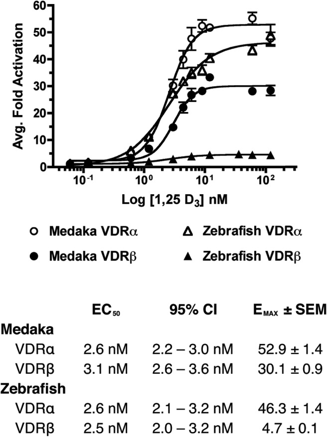
Concentration-response curves for the transactivation of medaka (circles) and zebrafish (triangles) VDRα (open) and VDRβ (closed) in response to 0–120 nM 1, 25 D3. HepG2 cells were transfected with pSG5-VDR, XREM-Luc, and pRL-CMV as an internal control as described in Materials and Methods. Data are represented as the average fold activation normalized to the vehicle control ± SEM (n = 4).