
Fig. 1.
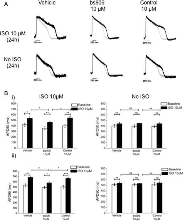
HSP20-PDE4D peptide disruptor modulates APD in hiPSC-CMs. hiPSC-CMs (Cellular Dynamics International, Madison, Wisconsin) were pre-treated for 2 h with vehicle (PBS), bs906 (10 μM) or control peptide (10 μM), followed by 24 h co-incubation with ISO (10 μM). Cells were loaded with 3 μM di-4-ANEPPS and electrical activity of spontaneously beating cardiomyocytes was registered using CellOPTIQ platform (Clyde Biosciences Ltd.). (A) Example AP traces (grey trace baseline, black trace 24 h 10 μM ISO/Vehicle). (B) (i) Average values of APD at 50% (APD50) and (ii) 90% of repolarisation (APD90) from baseline (white columns) or after treatment (black columns). Data represents mean ± S.E.M, measured from 15 areas per treatment from n = 3 separate cultures, ∗p < 0.05, ∗∗p < 0.01, ∗∗∗p < 0.001, one-way ANOVA post hoc Tukey’s test.