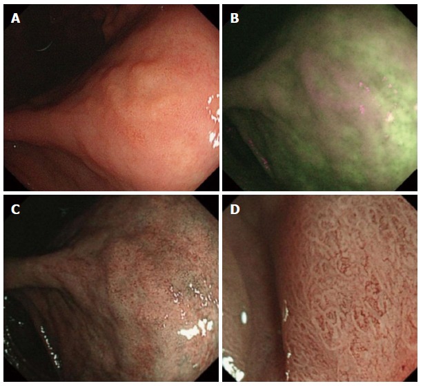
Figure 2.

Protruded early gastric cancer. A: Whitish lesion detected in white-light imaging; B: Magenta lesion in autofluorescence imaging; C: Demarcation line in narrow-band imaging (NBI); D: Irregular mucosal pattern and irregular vascular pattern in magnifying endoscopy with the NBI.
