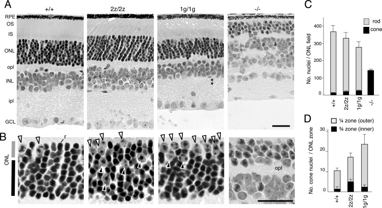
FIGURE 3.
Retinal morphology and gain of cones in mice lacking RORβ isoforms. A, histological sections revealed a normal retinal structure in RORβ2-deficient (2z/2z) mice but with an ∼2-fold excess of cone nuclei. RORβ1-deficient (1g/1g) mice also displayed excess cone nuclei and, as reported, loss of horizontal and amacrine cells and disorganized inner and outer plexiform layers. The asterisks indicate the missing amacrine cell layer in the inner nuclear layer. In comparison, Rorb−/− mice have almost exclusively cone-like cells instead of rods, lack photoreceptor segments, lack horizontal and amacrine cells, and display disorganized plexiform layers. RPE, retinal pigmented epithelium; OS, outer segment; IS, inner segment; opl, outer plexiform layer; INL, inner nuclear layer; ipl, inner plexiform layer; GCL, ganglion cell layer. B, higher magnification indicating cone nuclei (large with dispersed chromatin, arrowheads) and a representative rod nucleus (r, small with dense chromatin in +/+ mice (left panel)). Excess cone nuclei are present in 2z/2z and 1g/1g mice. Many cones in 2z/2z mice are misplaced in the inner zone of the ONL, but, in 1g/1g mice, most are in the outer zone. C, counts (mean ± S.D.) of cone and rod nuclei in 160-μm-long ONL fields showing excess cones in 2z/2z and 1g/1g genotypes (each p < 0.001 versus +/+) and an almost exclusive presence of cones in Rorb−/− mice. D, counts (mean ± S.D.) of cone nuclei in outer ¼ and inner ¾ zones of the ONL showing substantial dispersal of cones in the inner zone of 2z/2z mice (p < 0.001 versus +/+). Rorb−/− mice were not included in this comparison because they possess a thin ONL that cannot be compared with the ONL in the other mouse strains and because almost all cells in the ONL in Rorb−/− mice are cone-like. Scale bars = 25 μm.

