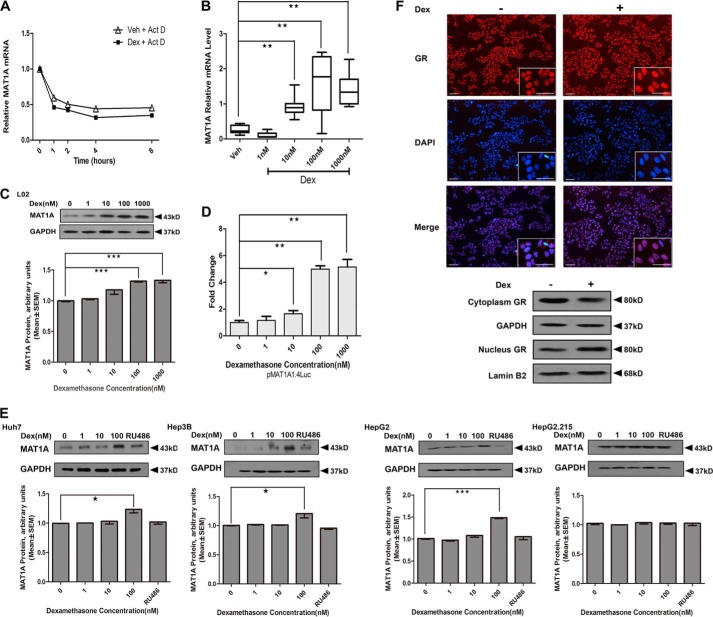
FIGURE 1.
Effect of Dex on MAT1A promoter activity and expression. A, analysis of MAT1A mRNA stability in L02 cells. Each level of Dex-treated and -untreated MAT1A mRNA before actinomycin D (Act D) treatment was considered as 1, and the relative levels were calculated. B and C, MAT1A mRNA and MAT1A protein were examined after L02 cells were treated with vehicle (Veh) or the indicated concentration of Dex for 24 h. D, effect of Dex concentration on the luciferase activity in L02 cells transfected with pMAT1A1.4Luc. E, MAT1A protein levels were detected in Huh7, Hep3B, HepG2 and HepG2.2.15 cells after treatment with the vehicle or Dex with or without RU486 for 24 h. The inset shows the representative immunoblots of different concentration points. *, p < 0.05; **, p < 0.01 and ***, p < 0.001. F, GR localization was investigated in the aforementioned cells treated with Dex for 12 h and then fixed, and endogenous GR was labeled (red). DNA was counterstained with DAPI (blue). GR protein levels and distributions were detected in the cytoplasm and nucleus, respectively. GAPDH or lamin B2 was used as a loading control. Scale bar, 50 μm. Shown is a representative of results from five independent experiments.

