
FIGURE 7.
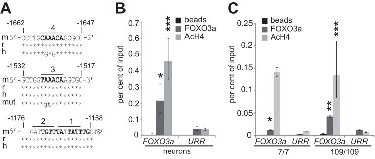
FOXO3a binds to its own promoter. A, in silico analysis of potential FHREs in mouse, rat, and human FOXO3a promoter. Shown are the conserved sequences of promoter regions containing the four identified sites that are marked in bold. The sequence of mutated FHRE 3 is also shown. The numbers indicate the positions relative to the transcription start site. B and C, chromatin immunoprecipitation analysis demonstrating FOXO3a binding to FHREs containing region in Foxo3a promoter in rat primary cortical neurons (B) and Hdh7/7 and Hdh109/109 cells (C). Soluble chromatin was co-immunoprecipitated with antibodies specific for FOXO3a or AcH4 or with beads alone. DNA from Foxo3a promoter or from an URR was amplified by qPCR. The data are presented as percentages of input DNA. The statistical significance denoted with asterisks is relative to values of URR obtained with the respective antibodies. *, p < 0.05; **, p < 0.01; ***, p < 0.001; n = 3 (except in case of AcH4 ChIP in Hdh7/7 cells, where n = 2).