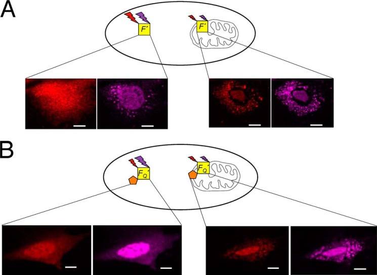
FIGURE 1.
A schematic diagram illustrating the utilization of a fluorescent internal reference to detect FRET between Alexa Fluor 546 and Dabcyl during translocation. A, the fluorescence intensities of Bax (F′) conjugated with the FRET donor (Alexa Fluor 546, red) and the internal reference (Alexa Fluor 633, purple) are measured during translocation. B, the fluorescence intensities of another Bax sample, FQ′, that contains the same FRET donor (red) and internal reference (purple) in addition to the non-fluorescent FRET acceptor (Dabcyl, orange) are also measured during translocation. Comparison of the relative intensities of the donor and internal reference with, FQ′, and without, F′, the acceptor under similar conditions would provide FRET efficiency. All scale bars are 10 μm.

