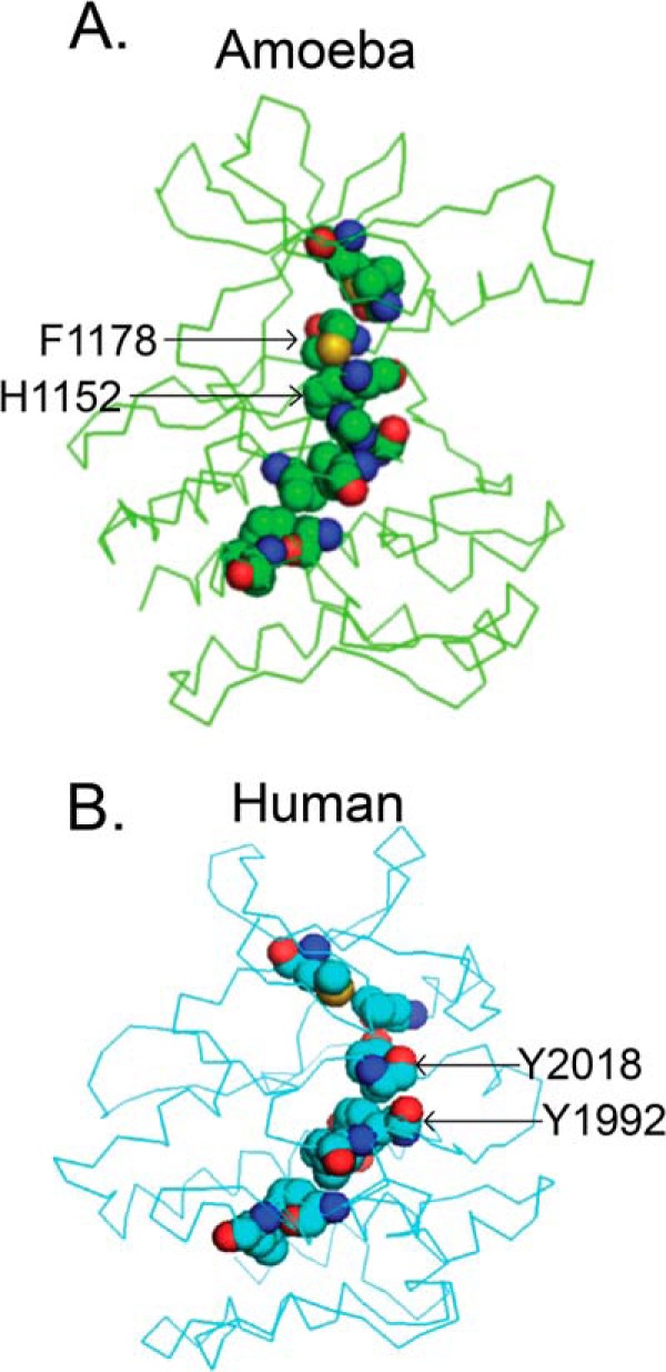
FIGURE 10.

Lack of conservation of the hydrophobic spine structure. Shown are the hydrophobic spines of ameba (A, green) and human LRRK2 kinase domain models (B, cyan). Residues involved in the hydrophobic spine are represented by spheres. Key residues on the conserved DXG and XRD motif are labeled by arrows. Elements are color-coded (light blue, carbon; red, oxygen; dark blue, nitrogen; yellow, sulfur).
