FIGURE 4.
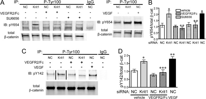
KRIT1 depletion leads to VEGF-dependent tyrosine phosphorylation of β-catenin. A, left panel, tyrosine 654 phosphorylation (pY654) of β-catenin in NC- and KRIT1-siRNA-transfected BAEC-treated ± 25 ng/ml VEGFR2/Fc or ± 1 μm SU6656. IgG, mouse IgG; IP, immunoprecipitation; IB, immunoblot. Right panel, comparison of KRIT1 siRNA-induced and VEGF-induced (50 ng/ml) Tyr-654 phosphorylation. Blots are representative, n = 4. B, densitometric quantification of β-catenin Tyr-654 phosphorylation from four independent experiments treated as in A. Tyr(P)-654 signal was normalized to β-catenin immunoprecipitation and displayed relative to NC-transfected cells, ±S.E. p = 0.002 by ANOVA; *, p < 0.01 by post-hoc testing versus NC-transfected cells; **, p < 0.05 versus KRIT1 siRNA-transfected cells. C, tyrosine 142 phosphorylation (pY142) of β-catenin in NC- and KRIT1-siRNA-transfected BAEC-treated ± 25 ng/ml VEGFR2/Fc or 50 ng/ml rhVEGF. The line indicates removal of intervening lanes. Blots are representative, n = 3. D, densitometric quantification of β-catenin Tyr-142 phosphorylation from three independent experiments treated as in C. The Tyr(P)-142 signal was normalized to β-catenin immunoprecipitation and displayed relative to NC-transfected cells ±S.E. p = 0.0003 by ANOVA; *, p < 0.05 by post-hoc testing versus NC-transfected cells; **, < 0.01 versus NC-transfected cells; ***, p < 0.05 versus KRIT1 siRNA-transfected cells.

