FIGURE 5.

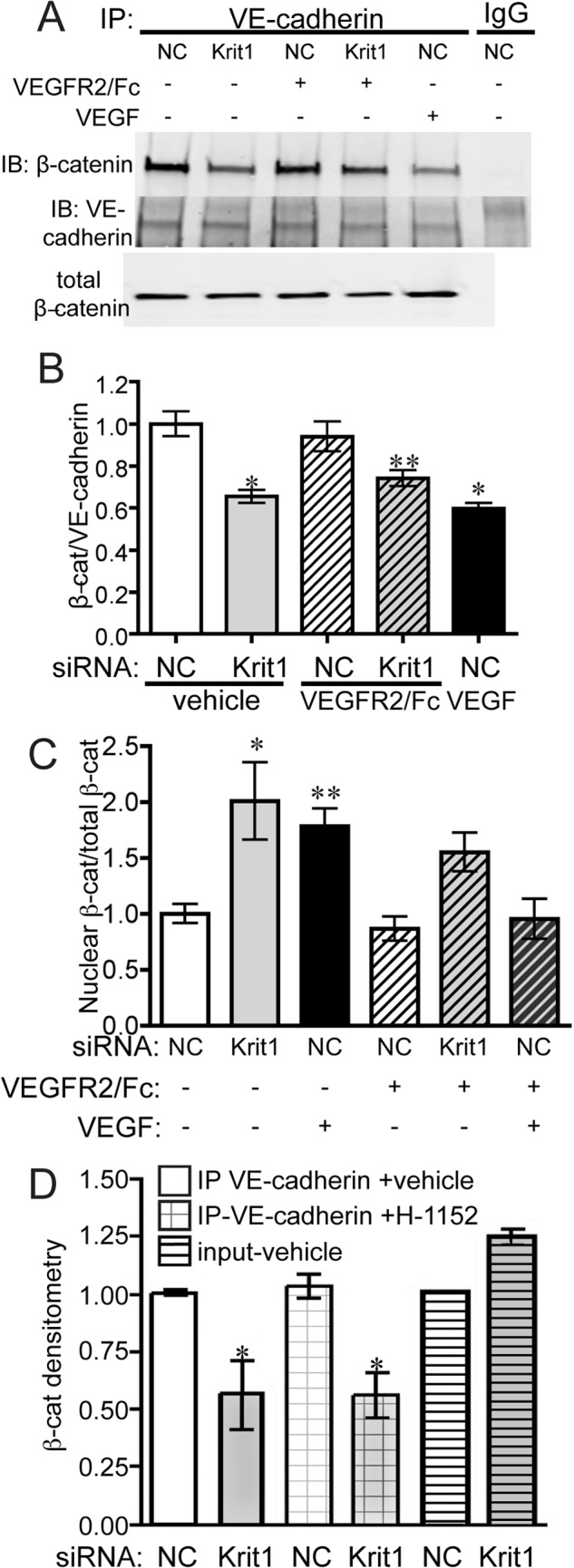
KRIT1 depletion-dependent dissociation of β-catenin and VE-cadherin is not reversed by inhibition of VEGFR2. A, co-immunoprecipitation of VE-cadherin and β-catenin in NC- and KRIT1-siRNA-transfected HPAEC-treated ± 25 ng/ml VEGFR2/Fc or rhVEGF (50 ng/ml). IgG, rabbit IgG; IP, immunoprecipitation; IB, immunoblot. Blots are representative, n = 5. B, densitometric quantification of β-catenin co-immunoprecipitation from five independent experiments treated as in A. Data shown are β-catenin-normalized to VE-cadherin immunoprecipitation ±S.E. p = 0.0004 by ANOVA; *, p < 0.001; **, p < 0.05 by post-hoc testing versus untreated NC-transfected cells. C, densitometric quantification of nuclear β-catenin in NC- and KRIT1-siRNA-transfected HPAEC-treated ± 50 ng/ml VEGFR2/Fc or rhVEGF (50 ng/ml). Nuclear fractions were isolated as described under “Experimental Procedures.” Data shown are nuclear β-catenin-normalized to total β-catenin for each condition from 5 independent experiments. p = 0.0013 by ANOVA; *, p < 0.01; **, p < 0.05 by Dunnett's multiple comparison test versus untreated NC-transfected cells. D, densitometric quantification of β-catenin co-immunoprecipitation with VE-cadherin in NC and KRIT1 siRNA-transfected HPAE treated with 100 nm H-1152. Data are combined from five independent experiments. Data shown are β-catenin-normalized to VE-cadherin immunoprecipitation ±S.E. p = 0.0082 by ANOVA; *, p < 0.05 by post-hoc testing versus vehicle-treated NC-transfected cells.
