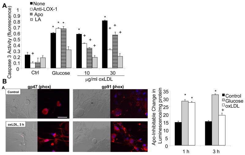
Fig. 5. High glucose and oxLDL cause cell death in DRG neurons via NAD(P)H Oxidase.
Adult DRG neurons were exposed to high glucose (25.7 mM) or increasing concentrations of oxLDL and then cell death was quantitated by caspase 3 activation after 5 h. DRG neurons were additionally pre-treated with LOX-1 neutralizing antibody (Anti-LOX-1, 100 mg/ml), apocyanin (Apo, 1 μM), or α-lipoic acid (LA, 100 μM). n=9, *p<0.01 compared to untreated control, +p<0.01 compared to no pre-treatment (None). (B) Adult DRG neurons were exposed to 30 μg/ml oxLDL and then immunolabeled for NAD(P)H oxidase subunits p47 or gp91. In (B), adult DRG neurons were exposed to high glucose (25.7 mM) or oxLDL (30 μg/ml) for 1 h or 3 h, then lysed for biochemical assays of NAD(P)H oxidase. *p<0.01 compared to untreated control, +p<0.05 compared to untreated control. (reproduced from (Vincent, et al., 2009b)).

