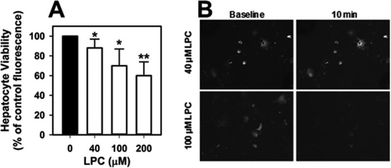
Fig. 8.
Concentration-dependent effects of LPC on hepatocyte viability. A. Primary mouse hepatocytes were incubated with 100 μM oleate complexed with albumin at a 5:1 ratio and LPC for 10 min prior to incubation with 2 μM calcein-AM and 8 mM CoCl2 at 37°C in the dark. Cell viability was determined based on cytoplasmic activity assessed by measuring fluorescence intensity after excitation at 485 nm and emission at 538 nm. The data represent mean ± standard deviations from 5 different experiments. * and ** denote differences from cells incubated without LPC at P < 0.05 and P < 0.01, respectively. B. Images of hepatocytes before and after incubation for 10 min with oleate/BSA in the presence or40 or 100 μM LPC. Intact mitochondria show bright punctate appearances.

