
Figure 4. Electrode placements in each experiment.
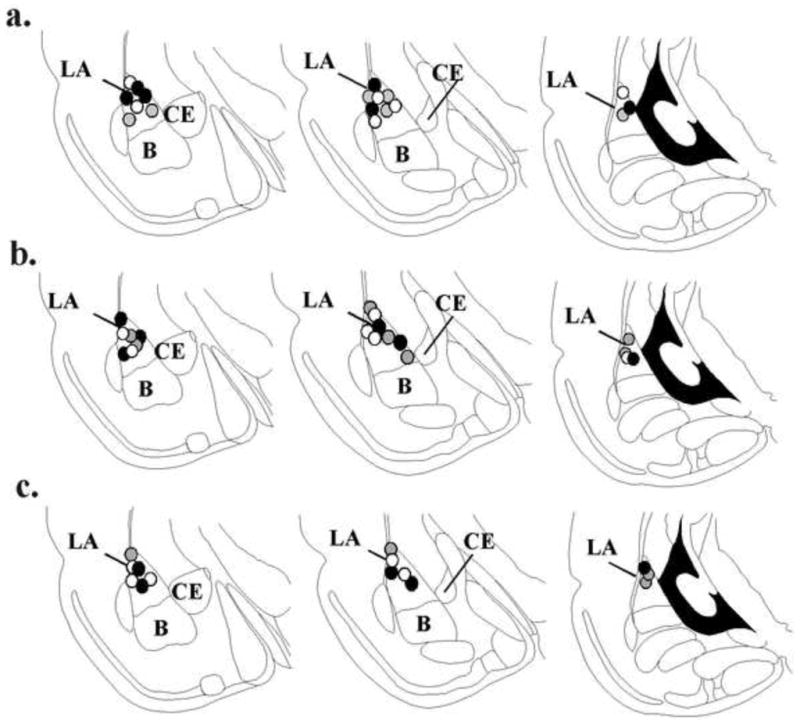
(a) Histological location of recording electrode placements for rats infused with vehicle (black circles), 5-AZA (gray circles) or RG108 (white circles) in the consolidation experiment (Figure 1). (b) Histological location of recording electrode placements for rats infused with vehicle (black circles), 5-AZA (gray circles) or RG108 (white circles) in the reconsolidation experiment (Figure 2). (c) Histological location of recording electrode placements for rats infused with vehicle (black circles), 5-AZA (gray circles) or RG108 (white circles) in the non-reactivated experiment (Figure 3). All panels adapted from Paxinos and Watson (1998). LA = lateral amygdala; CE = central nucleus of amygdala; B = basal nucleus of the amygdala