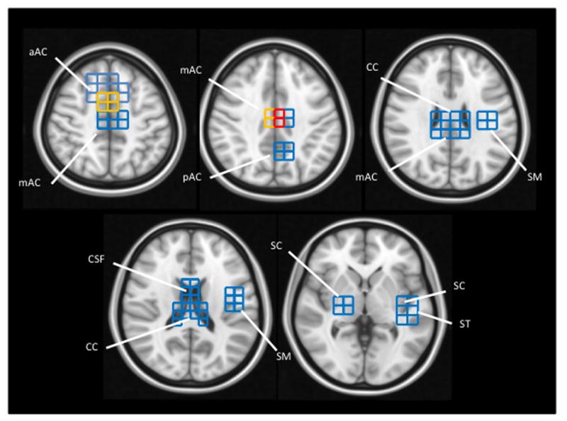
Figure 2. Lactate distribution in ASD participants.

Lactate-positive voxels found across all ASD participants are projected onto brain templates (downloaded from http://www.bic.mni.mcgill.ca/ServicesAtlases/ICBM152NLin2009). Voxel color indicates the number of ASD participants with a lactate doublet: Blue = 1 participant; Yellow = 2 participants; Red = 3 participants.
aAC: anterior portion of the anterior cingulate gyrus
mAC: mid-portion of the anterior cingulate gyrus
pAC: posterior portion of the anterior cingulate gyrus
CC: corpus callosum
CSF: cerebrospinal fluid
ST: superior temporal gyrus
SC: subcortical nuclei, including putamen, globus pallidus, thalamus (and associated internal capsule)
SM: sensory and motor portions of the pre- and post-central gyri
