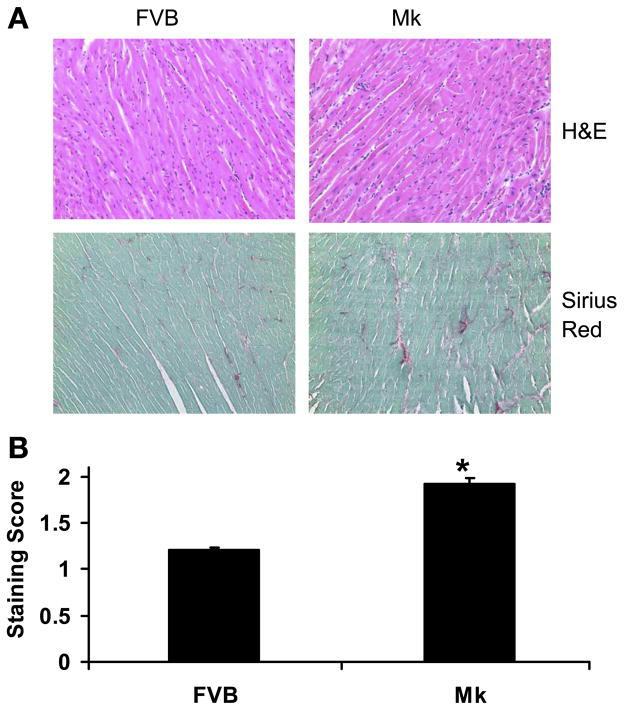
Fig. 8.
Fibrosis in Mk hearts. Cardiac morphology was visualized by hematoxylin and eosin (H&E) staining, and collagen accumulation was visualized by Sirius red staining at a magnification of ×40. A: representative staining of FVB and Mk hearts. B: average (±SE) scores for Sirius red staining from 60 photographs taken from 3 FVB and 3 Mk mouse hearts. Staining was rated by a blinded observer on a scale of 0–2, where 0 indicates mild interstitial accumulation of collagen, 1 indicates increased interstitial accumulation of collagen, and 2 severe interstitial accumulation of collagen. Values are means ± SE and were analyzed by Student’s t-test (*P < 0.01).

