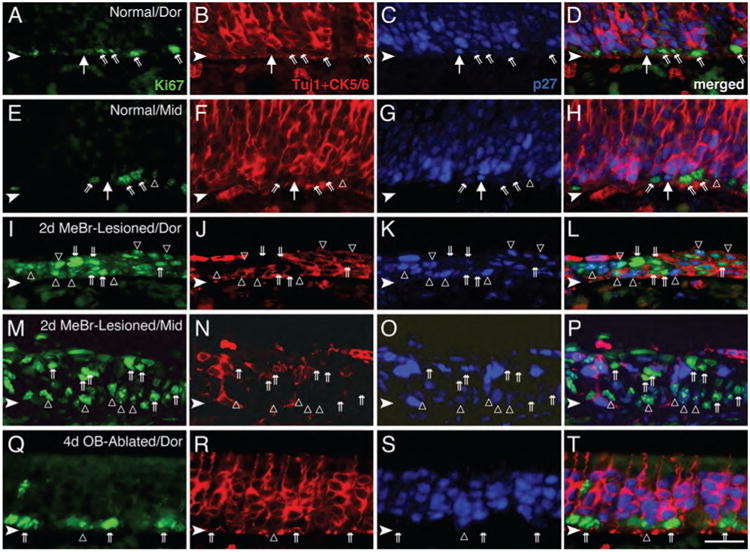
Figure 3.

Quiescent GBCs are present in the normal OE and disappear after MeBr lesion. A–H: The normal OE from dorsal (A–D) and mid-ventral (middle, E–H) septum was stained with anti-Ki-67 (green), anti-CK5/6 and Tuj-1 (red), and anti-p27Kip1 (blue). The GBC population is identified as those cells that label with neither Tuj-1 nor anti-CK5/6 and are located in the space between the CK5/6 (+) HBCs (deep to them) and Tuj-1 (+) neurons (just superficial to them). A large majority of the GBCs are Ki-67 (+) and p27Kip1 (−) (angled double arrows). However, some of the GBCs are p27Kip1 (+)/Ki-67 (−) (large arrows) and are presumed to be quiescent. Occasionally, cells that are p27Kip1 (+)/Ki-67 (+) are found (open triangles in E–H). I–P: At 2 days after MeBr lesion, the septal OE from dorsal (I–L) and ventral (M–P) regions show increased numbers of proliferating cells that are Ki-67 (+) (double arrows) compared with the normal OE (A–H), whereas p27Kip1 (+)-only cells are absent. Note that cells that are both p27Kip1 (+) and Ki-67 (+) (open triangles) are numerous after lesion. As described previously (Schwob et al., 1995), HBCs that are marked with anti-CK5/6 almost disappear in the ventral OE immediately after MeBr lesion (as in N). Q–T: At 4 days after olfactory bulb ablation, Ki-67 (+) dividing cells (double arrows) are more numerous than in normal epithelium, and cells labeled with p27Kip1 are less numerous and frequently also labeled with Ki-67 (open triangles). Abbreviations: Dor, dorsal septum; Mid, mid-point of septum. Arrowheads indicate the basal lamina. Magenta–green–blue versions of the photomicrographs are available online as Supplemental Figure S3. Scale bar = 25 μm in T (also applies to A–S).
