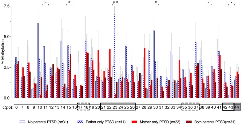
Figure 3.
Percent methylation at each of 39 C-phosphate-G dinucleotides (i.e., CpG sites) of the glucocorticoid receptor gene (NR3C1) exon 1F promoter (sites 6–43) and exon 1F region (site 44) analyzed by cytosine methylation bisulfite mapping (clone-based Sanger sequencing) using peripheral blood mononuclear cells (PBMCs) DNA, in a population of offspring described in our previous publications (Lehrner et al., 2014; Yehuda et al., 2014a). The 39 CpG sites accessed in the study are numbered as in Figure 1. The represented data (mean ± s.e.m.) are based on a multivariate Two-Way analysis of covariance with maternal and paternal post-traumatic stress disorder (PTSD) as fixed factors and parental Holocaust exposure, age, smoking history, and PBMC-type as covariates. μ, significant maternal PTSD effect. π, significant paternal PTSD effect. x, significant maternal by paternal PTSD interaction effect. Significance was set at p < 0.05. Boxes represent the location of known or putative canonical (solid-lined box) and non-canonical (broken-lined box) NGFI-A binding sites, according to McGowan et al. (2009), and a gray box represents the location of the beginning of exon 1F.

