Figure 3.
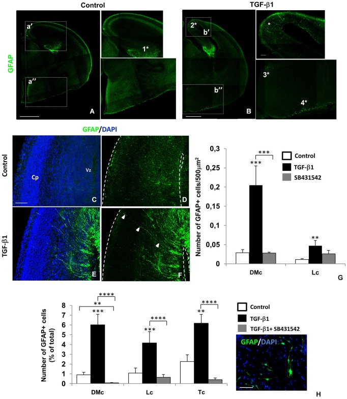
TGF-β1 promotes premature gliogenesis in the cerebral cortex. Intraventricular injection of TGF-β1 in mouse embryos (injection at E14 and analysis at E16) caused premature appearance of GFAP+ cells (green) in different telencephalon regions: dorsomedial cortex/cingulate cortex (2*), neuroepithelium related to the third ventricule (3*) and pial surface of the preoptic area (4*). At the hippocampal formation (1*), GFAP labeling was not affected. TGF-β1 induced gliogenesis was more evident at the dorsomedial area of the cerebral cortex (DMc), than in lateral cortex (Lc) (C–G). Note the GFAP+ (green) radial fibers of differentiating cells (arrows, F). In radial glia (RG) isolated cultures, TGF-β1 also promoted appearance of GFAP+ cells in a greater extend in DMc than in Lc and total cortex (Tc) (H). ***P < 0.0005, *P < 0.005. Scales: 500 μm (A,B), 50 μm (C,H). Cp: cortical plate, Vz: ventricular zone.

