Figure 1.

Hysterosalpingogram.
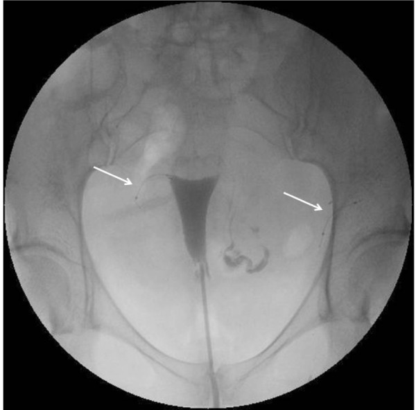
Notes: Hysterosalpingogram showing tubal occlusion on the right side with correct location of the device (right arrow) and a patent tube on the left side. The left device is abnormally positioned (left arrow).

Hysterosalpingogram.
Notes: Hysterosalpingogram showing tubal occlusion on the right side with correct location of the device (right arrow) and a patent tube on the left side. The left device is abnormally positioned (left arrow).